Webinar Nanos in Marseille
2021 september 10th – 2 to 4 PM
Registrations are free but mandatory at https://sfnanomarseille.sciencesconf.org

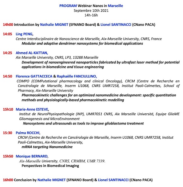
14h00. Introduction by Nathalie MIGNET (SFNano Board) et Lionel SANTINACCI (C’Nano PACA)
Les durées des présentations inclus les questions, compter 16-17 minutes maximum pour pouvoir répondre à quelques questions et laisser le temps d’installer les présentations
14:05 Ling Peng , Centre Interdisciplinaire de Nanoscience de Marseille, Aix-Marseille University, CNRS, France
Modular and adaptive dendrimer nanosystems for biomedical applications
14:25 Ahmed AL-KATTAN Aix Marseille University, CNRS, LP3, 13288 Marseille
Development of nanoengineered nanoparticles fabricated by ultrafast laser method for potential applications in biomedicine and tissue engineering
14:50 Florence GATTACCECA et Raphaëlle FANCIULLINO COMPO (COMPutational pharmacology and clinical Oncology), CRCM (Centre de Recherche en Cancérologie de Marseille, Inserm U1068, CNRS UMR7258, Institut Paoli-Calmettes, School of Pharmacy, Aix-Marseille University
Pharmacokinetic challenges for an optimized nanomedicine development: specific
quantitation methods and physiologically-based pharmacokinetic modelling
15h10 Marie-Anne Estève, Institut de NeuroPhysiopathologie (INP), UMR7051 CNRS, Aix Marseille Université, Equipe GlioME Gliomagenesis and MicroEnvironment
Nanosystems and ultrasounds as tools to improve glioblastoma treatment
15:30 Palma ROCCHI, CRCM, Centre de Recherche en Cancérologie de Marseille, équipe Nanoparticules et ciblage thérapeutique
mRNA targeting Nanomedicine
15h50 Monique Bernard, Center for Magnetic Resonance in Biology and Medicine
Perspectives en imagerie
16h00 Mots de la fin by Nathalie MIGNET (SFNano Board) et Lionel SANTINACCI (C’Nano PACA)
16h05 End of the webinar
Dr Ling Peng is a CNRS research director at Centre Interdisciplinaire de Nanoscience de Marseille. She is a leading expert in developing functional dendrimer nanosystems for biomedical applications. She has successfully established bio-inspired and self-assembling dendrimer nanosystems for the delivery of imaging agent, drug and nucleic acid therapeutics for cancer treatment and diagnosis. Her team has been labelled by La Ligue Contre Le Cancer for developing nanotheranostics in cancer therapy. Dr Peng is a distinguished member of French Chemical Society, and was awarded by the French Academy of Science with the Prize of Dr & Mrs Henri Labbé.
 Modular and adaptive dendrimer nanosystems for biomedical applications
Ling Peng
Centre Interdisciplinaire de Nanoscience de Marseille, Aix-Marseille University, CNRS, France
Email : ling.peng@univ-amu.fr
The application of nanotechnology is widely expected to bring breakthrough in medicine for disease treatment and diagnosis. Dendrimers are ideal materials for elaborating nanomedicine by virtue of their well-defined structure, multivalent cooperativity and nanosize per se. We have pioneered modular and adaptive self-assembling dendrimer nanosystems for the delivery of imaging agents, drugs and nucleic acid therapeutics.1 Benefiting from the multivalent feature of dendrimer and the EPR effect of the tumor microenvironment, these dendrimer nanosystems offer excellent tumor imaging, outperforming the clinical imaging references.2 In addition, they penetrate deep in tumor tissue and bypass drug efflux, effectively overcoming drug resistance.3 Also remarkably, these nanosystems deliver nucleic acid therapeutics effectively in primary immune cells, offering great promise in immunotherapy.4
- Acc. Chem. Res. 2020, 53, 2936.
- Proc. Natl. Acad. Sci. U.S.A. 2018, 115, 11454; Small 2020, 16, 2003290; Chem. Commun. 2020, 56, 301.
- Proc. Natl. Acad. Sci. U.S.A. 2015, 112, 2978; Exploration 2021, DOI: 10.1002/EXP.20210003
- Nat. Protoc. 2021, 16, 327; Nat. Commun. 2020, 11, 1773; J. Am. Chem. Soc. 2018, 140, 16264; Angew. Chem. Int. Ed. 2014, 53, 11822.
Development of nanoengineered nanoparticles fabricated by ultrafast laser method for potential applications in biomedicine and tissue engineering
Ahmed Al-Kattan
Aix Marseille University, CNRS, LP3, 13288 Marseille
ahmed.al-kattan@univ-amu.fr
Driven by surface cleanness, unique physical and optical and chemical properties, bare(ligand-free) laser synthesized nanoparticles (NPs) are now in the focus of intensive researches for varieties of applications such environment, catalysis and biomedicine [1]. Based on the interaction of ultra-pulsed laser beam in liquid ambiance (e.g., aqueous solution) with a solid target material or water powder dispersed, this process can lead naturally to the formation of spherical NPs with modulate physicochemical properties including diameter and size dispersion, surface chemistry free from any ligands and functionalization. For instance, we have thus demonstrated the possibility to elaborate ultraclean and extremely stable colloidal solutions based on variety of composition including of SiNPs, AuNPs, Si@AuNPs or even TiNNPs with flexible physicochemical properties [1]. Moreover, the presence of high oxidation state on the NPs surface promises efficient interactions with many biological materials (e.g., proteins), and other chemical functions [1]. We have shown that such NPs can be exploited as significant sensitizers for radiofrequency (RF)-induced hyperthermia, photothermal or even TPE-PDT therapeutic modalities [2-4]. Beside conventional additives mainly made-it by chemical way, we have also started to explore such bare laser-synthesized NPs as novel functional additives for tissue engineering applications [5].
[1] A. Al-Kattan, D. Grojo, C. Drouet, A. Mouskeftaras, P. Delaporte, A Casanova, J. D. Robin, F. Magdinier, P. Alloncle, C. Constantinescu, V. Motto-Ros and J. Hermann, Short-pulse lasers: a versatile tool in creating novel nano-/micro-structures and compositional-analysis for healthcare and wellbeing challenges. Nanomaterials, 11(3), 712, 2021
[2] K. P. Tamarov, L. A. Osminkina, S. V. Zinovyev, K. A. Maximova, J. V. Kargina, M. B. Gongalsky, Y. Ryabchikov, A. Al-Kattan, A. P. Sviridov, M. Sentis, A. V. Ivanov, V. N. Nikiforov, A. V. Kabashin and Victor Yu Timoshenko, Radio frequency radiation-induced hyperthermia using Si nanoparticle-based sensitizers for mild cancer therapy, Sci. Rep, 4, 7034, 2014
[3] A. A. Popov, G. Tselikov, N. Dumas, C. Berard, K. Metwally, N. Jones, A. Al-Kattan, B. Larrat, D. Braguer, S. Mensah, A. Da Silva, M.-A. Estève and A. V. Kabashin, Laser- synthesized TiN nanoparticles as promising plasmonic alternative for biomedical applications, Sci. Rep., 9, 1194, 2018
[4] A. Al-Kattan, L. M.A. Ali, M. Daurat, E. Mattana and M. Gary-Bobo, Biological assessment of laser-synthesized Silicon nanoparticles effect in two-photon photodynamic therapy on breast cancer MCF-7 cells, 10(8),1462, 2020
[5] A. Al-Kattan, V. P. Nirwan, A. A. Popov, Y. V. Ryabchikov, G. Tselikov, M. Sentis, A. Fahmi, A. V. Kabashin, Recent advances in laser-ablative synthesis of bare Au and Si NPs and assessment of their prospects for tissue engineering applications, Int. J. Mol. Sci., 19, 1563, 2018

Ahmed Al-Kattan is Assoc. Prof. at LP3 laboratory (UMR 7341,CNRS-Aix-Marseille University). His research is dealing with the design of novel biocompatible/biodegradable nanoparticles fabricated by ultrafast laser process and soft chemistry for advanced theranostic modalities, and the development of innovative functional biomimetic scaffold platforms for tissue engineering applications.


Pharmacokinetic challenges for an optimized nanomedicine development: specific quantitation methods and physiologically-based pharmacokinetic modelling.
Florence Gattacceca, PharmD, PhD, HDR, Raphaëlle Fanciullino, PharmD, PhD, HD
COMPO (COMPutational pharmacology and clinical Oncology), CRCM (Centre de Recherche en Cancérologie de Marseille, Inserm U1068, CNRS UMR7258, Institut Paoli-Calmettes,School of Pharmacy, Aix-Marseille University
Résumé
The promise of nanomedicines is to provide improved pharmacokinetic (PK) properties compared to conventional formulations. However, knowledge on the influence of the characteristics of nanomedicines on their behaviour in the organism is still limited. To fill this gap, accurate quantitation methods and integrated analysis of collected in vitro and in vivo PK data are needed.
The importance of a specific quantitation of the free versus nanoformulation-encapsulated drug will be underlined and exemplified based on Vyxeos example (R Fanciullino). How physiologically-based pharmacokinetic models are an ideal platform to integrate heterogeneous information will be illustrated (F Gattacceca).
Dr Raphaelle Fanciullino, Pharm.D. Ph.D., is Assistant Professor of Pharmacokinetics and Clinical Pharmacist at Aix Marseille University and the University Hospital of Marseille, France. Dr Fanciullino work focus on controlling the pharmacokinetics variability of anticancer agents using either pharmacogenetics testing or developing drug delivery systems. Dr Fanciullino has prototyped a variety of stealth liposomes and immunoliposomes encapsulating several cytotoxics in colorectal cancer and breast cancer models. Dr Fanciullino has PI’ed several clinical trials in the field of clinical pharmacokinetics, and supervised and mentored several Ph.D students. Dr Fanciullino has published 50 international articles in oncology to date, and owns several patents on original liposomal formulations in oncology.
Dr Florence Gattacceca, pharmacokinetics expert, associate professor in pharmacokinetics at Aix-Marseille University since 2017, assistant professor in pharmacokinetics at Montpellier University from 2007 to 2017, received her PhD in Toxicology from University Paris-Est Créteil in 2004.Co-author of 26 peer-reviewed scientific publications (h-index 12). She was a visiting scientist at Northeastern University, Boston, USA, in Pr Amiji’s lab, in 2012-2013. Board member (2016-2021), secretary (2019) and president (2020) of GMP (Group of Metabolism and Pharmacokinetics). Pharmacokinetics expert at ANSM (French national drugs agency) since 2016. Member of the COMPO (Computation pharmacology and clinical oncology) team. Her research focuses on treatment optimization and individualization through pharmacometrics, in clinical practice and for the development of small molecules and nanomedicines.

Nanosystems and ultrasounds as tools to improve glioblastoma treatment
Marie-Anne Estève
Institut de NeuroPhysiopathologie (INP), UMR7051 CNRS, Aix Marseille Université
Equipe GlioME Gliomagenesis and MicroEnvironment
Central nervous system (CNS) diseases are among the most difficult to treat due to the presence of the blood brain barrier (BBB). As an example, the median survival of patients with glioblastoma, the most aggressive CNS tumor, is only 15 months despite standard treatment protocols combining surgery, chemotherapy and radiation. Recent strategies have been developed for delivering therapeutics across the BBB. One of them is the development of efficient nano-sized drug delivery systems (DDS) able to cross the BBB. Another one is the use of focused ultrasound-mediated BBB opening (FUS-BBBop), combined with microbubbles, as a non-invasive strategy to permeabilize the BBB locally, temporary, and reversibly (Marty et al, 2012). In this context, in the BubDrop4Glio transdisciplinary project, our aim is to develop perfluorocarbon nanoemulsions to encapsulate a hydrophobic drug (e.g., docetaxel) as sonosensitive DDS and to combine them to FUS-BBBop to increase drug concentration at tumor site. Other projects developed in our group combine FUS-BBBop to photothermal therapy for glioblastoma, using a multifunctional preclinical device developed for this purpose.
Dr Marie-Anne Estève is assistant professor and hospital pharmacist at Aix Marseille University, Assistance Publique – Hôpitaux de Marseille (Marseille, France). She carries out her research activity at the Institute of NeuroPhysiopathologie (CNRS, UMR7051), in the Gliomagenesis and Microenvironment (GlioME) team. Her earlier works focused on pharmacological and preclinical studies of anticancer drugs, with a particular interest on chemotherapeutic drugs targeting microtubules (e.g., Vinca-alkaloids and taxanes). In the last ten years, she used nanocarriers as drug delivery systems to overcome glioblastoma challenges, through multidisciplinary projects. The aim of her group is to investigate the safety, anticancer efficacy, pharmacokinetics and biodistribution profile of different nanoparticles (metallic nanoparticles, carbon-based nanoparticles, nanodroplets) using adapted in vitro and in vivo models.

Palma Rocchi, PhD-HDR
Centre de Recherche sur le Cancer, Marseille
Prostate Cancer Nanomedicine Group Leader
Dr Palma Rocchi received her PhD in 2002 from the Medicine Faculty of Marseille evaluating the molecular mechanisms involved in the castration-resistant prostate cancer (CRPC). In her three years as post-doctoral fellow in the University of British Columbia Prostate Centre in Dr Gleave’s Lab. Palma Rocchi completed her formation by receiving advance training in targeting mRNA of genes associated with CR and treatment resistance (TR) in PC. She developed a drug (OGX-427) against Hsp27 mRNA using oligonucleotide antisens (OA) and small interference RNA (siRNA) approach able to significantly dose-dependent down-regulate Hsp27 protein expression level. OGX-427 has been patented and obtained an international license (Apatorsen) by the University of British Columbia and Oncogenex and clinical trials phase II was done in Canada and United States to evaluate the effect of OGX-427 in patients with PC and other solid tumors like breast, ovarian, bladder, prostate and lung cancer. She has now completed her post-doctoral fellowship and found a position as an independent senior investigator in PC translational research at the French National Institute for Health and Medical Research (Inserm). She now holds a research director position working in prostate cancer translational research at the French National Institute for Health and Medical Research (Inserm). She focuses on oligonucleotide-based RNA targeting nanotherapies to improve treatment in PC and other diseases, in the setting of many national and international collaborations.
mRNA targeting Nanomedicine
Résumé
Antisense oligonucleotides (also called ASOs) belong to the family of therapeutic oligonucleotides. ASOs are used to perform selective silencing of mRNAs with subsequent knockdown of proteins (for coding mRNA) either at in vitro or in vivo scale. Generally, ASOs carry out their functions by base pairing in the cytoplasm or the nucleus of cells. The single stranded DNA ASO specifically binds to its complementary strand on target mRNA and forms an hetero duplex that recruits an active enzyme complex. This enzyme cleaves the target mRNA or prevent ribosome assembly machinery, leading in the both cases to the inhibition of translation. The main drawback of ASOs relies on their poor cellular penetration. ASOs are therefore usually delivered via transfecting reagent which could be cytotoxic. Therefore, improvement of ASO delivery is of major importance for in vitro and in vivo applications. To circumvent this issue, we have developed an original approach based on lipidic-ASO called LASO which have the property for self-assembling into nanomicelles allowing their internalization without using any transfecting reagent. This has been proved in prostate cancer (PC) models (Karaki, S., JCR, 2017) but can be used as nanomedicine to treat different diseases.

Dr Monique Bernard is research director at CNRS and is heading CRMBM (Centre de Résonance Magnétique Biologique et Médicale, CNRS and Aix Marseille Université) since 2012 where she is co-head of the cardiovascular group. Her research interests are focused on the study of metabolic, physiological and functional alterations in cardiovascular pathologies using magnetic resonance imaging (MRI) and spectroscopy (MRS, 31P, 23Na and 1H). She has published 155 original papers and reviews, and 15 book chapters. She is coordinator of the Marseille hub of France Life Imaging, deputy director of Marseille Imaging Institute at Aix-Marseille University.
Perspectives in biomedical imaging
Monique Bernard
Aix-Marseille Univ, CNRS, CRMBM, UMR 7339, Marseille.
In the era of precision medicine and the development of imaging methods to monitor disease progression and stratify patients to therapies that will benefit them, there is a growing need for specific molecular information. However, imaging techniques often suffer from a lack of target specificity and lesion location information. The development of imaging agents, contrast agents or radiopharmaceuticals, depending on the imaging modality, is therefore of great interest to provide this information. In addition, theranostic approaches that combine diagnosis and therapy are very promising. This is true for all imaging modalities, whether alone or combined in multi-modality imaging, where nanoparticles are receiving much attention. However numerous nanoparticles are developed and the path to biomedical applications and clinical implementation is long and difficult. From the development of nanoparticles, it is important to keep the end use in mind. Chemists, biologists, physicists and physicians have to work together to optimally integrate the development of nanotechnology and imaging techniques and to strategically combine the efforts.


