SFNano is pleased to announce the first webinar dedicated to promising young scientists.
100 participants were present to listen the young researchers: THANK YOU !
You miss it ? The webinar is now accessible online here !
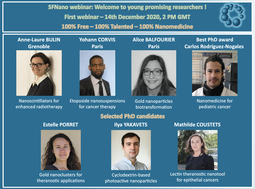
Monday 14th December, you will be able to listen:
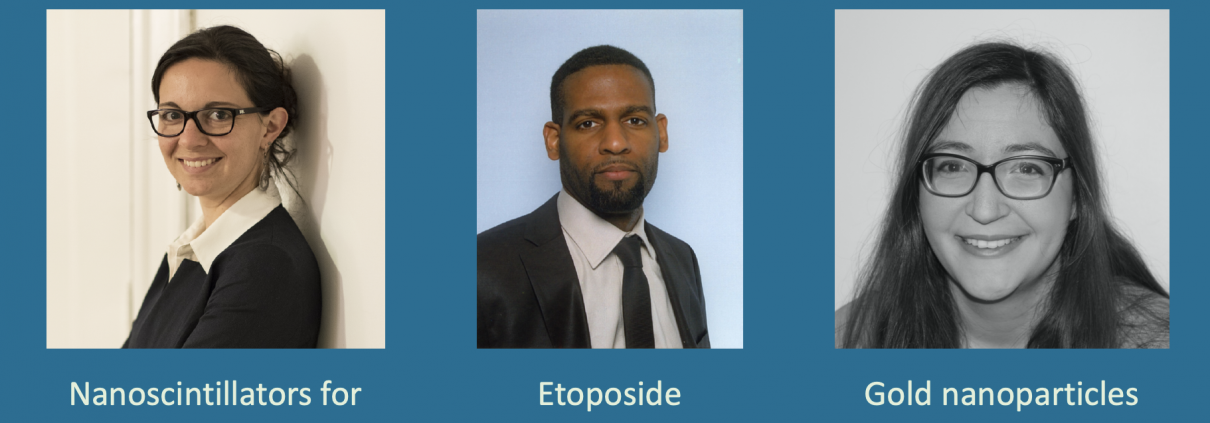
- Dr Anne-Laure BULIN, Synchrotron Radiation for Biomedicine, INSERM UA7 – Université Grenoble Alpes: Nanoscintillators for enhanced radiotherapy.
To improve the prognosis of glioblastoma, innovative radiotherapy regimens are required to augment the effect of tolerable radiation doses while sparing surrounding tissues. In this context, nanoscintillators are emerging radiotherapeutics that down-convert X-rays into photons with energies ranging from UV to near-infrared. As they contain high-Z elements, nanoscintillators are likely to induce a radiation dose-enhancement effect upon X-ray irradiation. This phenomenon stems from a higher photoelectric absorption of orthovoltage X-rays by high-Z elements compared to tissues, resulting in increased production of tissue-damaging photo- and Auger electrons. In this study, Geant4 simulations reveal that rare-earth composite LaF3:Ce nanoscintillators effectively generate photo- and Auger-electrons upon orthovoltage X-rays. 3D spatially resolved X-ray fluorescence microtomography shows that LaF3:Ce highly concentrates in microtumors and enhances radiotherapy in an X-ray energy-dependent manner. In an aggressive syngeneic model of orthotopic glioblastoma, intracerebral injection of LaF3:Ce is well tolerated and achieves complete tumor remission in 15% of the subjects receiving monochromatic synchrotron radiotherapy. This study provides unequivocal evidence for radiation dose-enhancement by nanoscintillators, eliciting a prominent radiotherapeutic effect. Altogether, nanoscintillators have invaluable properties for enhancing the focal damage of radiotherapy in glioblastoma and other radioresistant cancers. 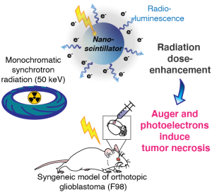
- Dr Yohann CORVIS, UTCBS, Unité de Technologies Chimiques et Biologiques pour la Santé, CNRS UMR 8258 – Inserm U 1267, Paris: Etoposide nanosuspensions for cancer therapy.
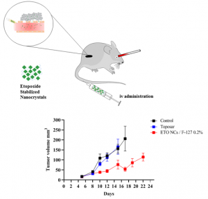
Etoposide, which is largely used as an anti-cancerous agent against testicular, lymphomas, ovarian, small cell lung, colon and breast cancer in its liquid dosage form, has been selected to design and engineer injectable nanocrystal suspensions with high benefit-risk ratio properties. We provide first and foremost optimized formulations for nanostructured etoposide dispersed solutions and validate by means of in vitro and in vivo evaluations the efficiency of this multiphase system. Indeed, the etoposide formulated as nanosuspension by a bottom-up approach, showed higher blood life span, reduced tumor growth and higher tolerance in murine carcinoma cancer models. The results obtained are promising for future clinical evaluation of these etoposide nanosuspensions. This research work was developed from the state of the art pointing out the fact that no antitumoral drug was marketed as nanocrystal i.v. dosage form. The corresponding review article was published in ChemMedChem by our group in early 2019; with two distinctions: i) ChemMedChem Cover Feature, and ii) One of the top ChemMedChem articles gathered in « ChemMedChem Hot Topic » Special Collection Series in February 2020 to celebrate World Cancer Day and raise awareness about cancer and promote understanding and further research regarding its detection, treatment, and prevention. Furthermore, the nanocrystals formulation protocol has been patented in 2018 (WO 2020043735). The original work presented could be of interest both to a large general public and to the researchers who work on administration of nanoparticles suspensions of drugs for targeting optimization, sustained release application, and overall therapeutic efficiency.
- Dr Alice Balfourier, laboratoire Matière et Systèmes Complexes, Université de Paris, Paris: Gold nanoparticle biotransformation.
Gold nanoparticles are promising devices for nanomedicine, for imaging, therapy, diagnosis or vectorization. However, due to their chemical inertia, their fate in the body remains little known, especially at the cellular level. We observed the biotransformation of gold nanoparticles of two sizes (4 and 20 nm in diameter) in cultured fibroblasts for several months using electron microscopy, and thus highlighted an unexpected phenomenon: the degradation of gold nanoparticles in lysosomes. This phenomenon leads to a recrystallization of the gold in an original form of sheets composed of self-organized clusters. We also shed light on the chemical and biological species involved in this transformation by studying the transcriptomic response of the cell to gold nanoparticles. These results open new perspectives for the use of gold, in nanoparticle or ionic form, in medicine.

In addition, selected young researchers will present their work evaluated for the best nanomedicine PhD 2020.
The web link will be provide soon. Don’t miss the SFNano webinar, it’s free to all !

